Like a canary in a coal mine, bleached coral serves as nature’s alarm system for ocean health crisis. You’ll observe the transformation as vibrant reef ecosystems lose their symbiotic zooxanthellae, exposing stark white calcium carbonate skeletons beneath. Temperature increases of just 1-2°C above seasonal maximums trigger this expulsion process within 48-72 hours. Understanding the precise visual indicators—from initial pallor to complete tissue whitening—becomes critical when coral mortality rates reach 50% within weeks of onset.
The Science Behind Coral Bleaching: Understanding the Process
When water temperatures rise just 1-2°C above normal seasonal maximums, you’re witnessing the trigger for one of the ocean’s most devastating biological processes.
You’re observing the breakdown of a vital symbiotic relationship between coral polyps and zooxanthellae algae. These microscopic algae provide up to 90% of the coral’s energy through photosynthesis, living within the coral’s tissues in exchange for nutrients and protection.
Heat stress disrupts this partnership. You’ll see corals expelling millions of zooxanthellae cells per square centimeter within hours.
Ocean acidification, pollution, and ultraviolet radiation accelerate this expulsion. Without their algal partners, corals lose their primary food source and vibrant coloration.
They’re left transparent, revealing their white calcium carbonate skeletons underneath—the characteristic “bleached” appearance you’ll recognize. Additionally, ocean acidification leads to difficulty in coral calcium carbonate skeleton production, further exacerbating the challenges faced by these vital ecosystems.
Visual Stages of Coral Bleaching: From Healthy to Severely Stressed
Although coral bleaching occurs rapidly, you’ll observe distinct visual stages that progress from subtle stress indicators to complete whitening over days or weeks.
Stage 1: Initial Stress Response
You’ll notice slight color dulling and reduced fluorescence as zooxanthellae begin expelling. Coral polyps may contract more frequently, and tissue appears less vibrant than baseline conditions.
Stage 2: Partial Bleaching
You’ll observe patchy white areas developing across 20-50% of coral surfaces. Coloration becomes mottled with distinct boundaries between stressed and healthy tissue sections.
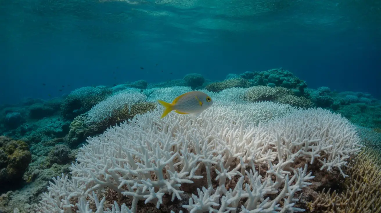
Stage 3: Severe Bleaching
You’ll witness 70-100% tissue whitening as symbiotic algae populations drop below 1 million cells per square centimeter. Coral skeletons become visible through transparent tissue, creating stark white appearances that indicate critical stress levels requiring immediate environmental intervention. Healthy reefs are essential for supporting biodiversity and maintaining ecosystem balance, making their preservation crucial in the face of such stressors.
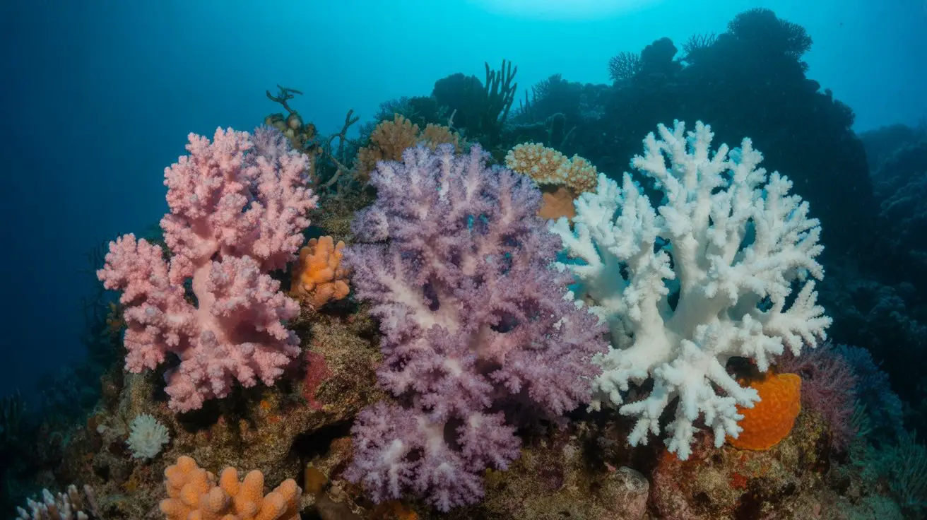
Healthy Coral vs. Bleached Coral: Key Visual Differences
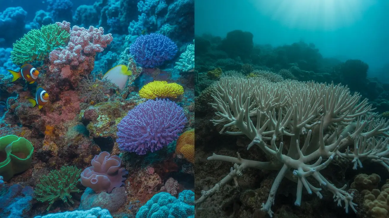
Understanding these bleaching progression stages becomes clearer when you compare specific characteristics between healthy and compromised coral systems.
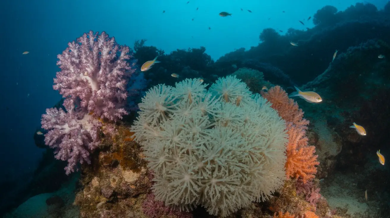
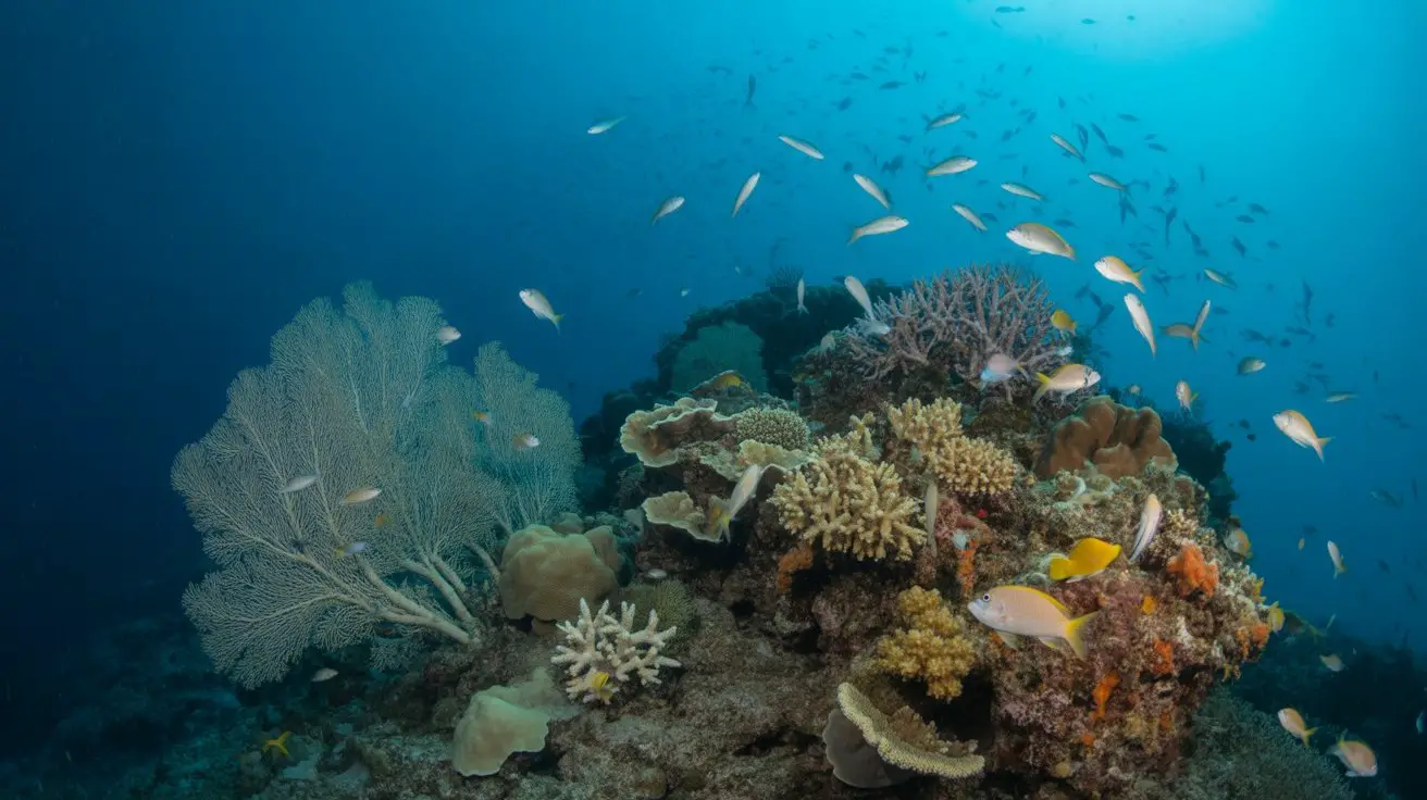
Healthy corals display vibrant pigmentation through symbiotic zooxanthellae, creating browns, greens, yellows, and reds with consistent coloration across polyp structures. You’ll observe active polyp extension during feeding periods and normal tissue thickness covering the calcium carbonate skeleton.
Bleached corals exhibit stark white or pale coloration as zooxanthellae densities drop from normal levels of 1-2 million cells per square centimeter to under 500,000.
You’ll notice translucent tissue revealing the underlying white skeleton, reduced polyp activity, and increased susceptibility to algal overgrowth.
Advanced bleaching shows tissue recession, exposing bare skeleton areas, and compromised structural integrity with visible deterioration patterns. Additionally, the health of coral reefs is crucial for biodiversity ecosystems, as they support numerous marine species and contribute to overall ocean health.
Early Warning Signs: Spotting Coral Stress Before Complete Bleaching
Before corals reach complete bleaching, you’ll detect subtle physiological changes that signal mounting stress within the symbiotic relationship. These precursor indicators manifest as measurable alterations in coral coloration, behavior, and tissue integrity before zooxanthellae expulsion occurs.
Monitor these critical stress indicators:
- Pale coloration patterns – Corals exhibit reduced pigmentation intensity, appearing 20-30% lighter than baseline coloration while retaining visible zooxanthellae populations.
- Tissue recession – Polyp retraction increases during daylight hours, with skeletal exposure becoming visible at colony margins and growth tips.
- Reduced feeding response – Diminished capture rates of prey items and decreased tentacle extension during ideal feeding periods.
You’ll observe these changes 2-4 weeks before complete bleaching events. Document color variations using standardized coral health charts and photograph colonies under consistent lighting conditions for accurate stress assessment. Additionally, understanding the impact of coral reef ecosystems can provide valuable insights into the health of marine environments.
Different Coral Species and Their Bleaching Patterns
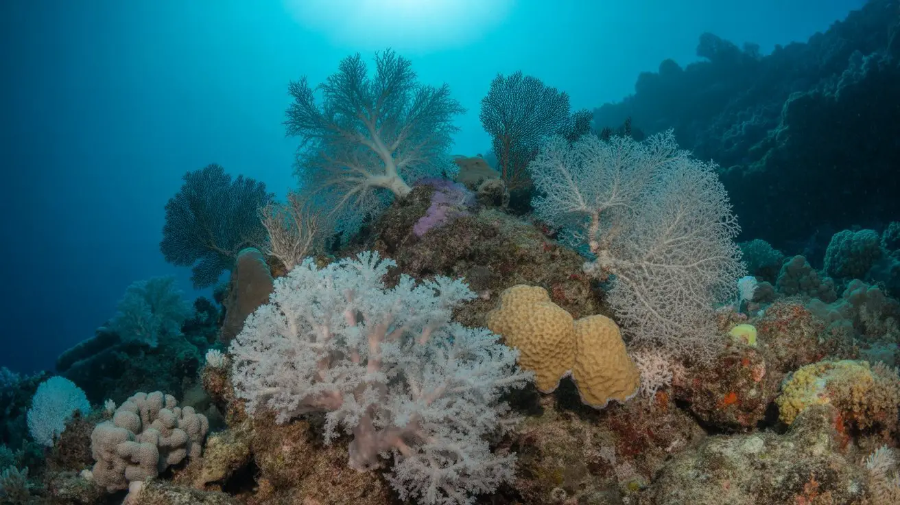
Coral species exhibit distinct bleaching patterns that reflect their unique physiological adaptations and symbiotic relationships with specific zooxanthellae clades.
You’ll observe that branching corals like Acropora bleach rapidly, losing color within 48-72 hours of thermal stress exceeding 1°C above maximum monthly mean temperatures.
Massive corals such as Porites resist bleaching longer, maintaining partial pigmentation for 2-3 weeks under identical conditions.
You’ll notice that plate corals like Turbinaria show patchy bleaching, retaining zooxanthellae in shaded areas while surface tissues whiten completely.
Brain corals exhibit sectoral bleaching, where individual polyps bleach while adjacent ones remain colored.
You can document that soft corals respond differently—they often retract tissues rather than expel symbionts immediately, appearing deflated before color loss occurs. Additionally, the health of coral reefs is influenced by predator populations, which help maintain the balance of the ecosystem and can impact coral resilience during bleaching events.
Environmental Factors That Trigger Coral Bleaching Events
When ocean temperatures rise just 1-2°C above the long-term summer maximum for 4-6 weeks, you’ll witness the primary trigger for mass bleaching events across reef systems.
However, thermal stress isn’t the sole culprit behind these devastating phenomena.
Multiple environmental stressors can initiate bleaching responses:
- Ocean acidification – pH levels dropping below 7.8 disrupt symbiotic algae metabolism, weakening coral resilience to temperature fluctuations.
- Solar irradiance intensity – UV radiation exceeding 400 watts/m² during low tide exposure compounds thermal stress effects.
- Pollution runoff – Nitrogen concentrations above 1.0 μM and sediment loads create additional physiological stress on coral polyps.
You’ll observe that these factors often interact synergistically, meaning combined stressors trigger bleaching at lower individual thresholds than isolated conditions would typically require.
Additionally, the impact of temperature variations on coral health underscores the urgency in addressing climate change to protect these vital ecosystems.
Recovery Signs: How to Identify Corals Bouncing Back From Bleaching
You’ll identify recovering corals through active polyp extension during nighttime feeding periods and resumed calcium carbonate secretion, evidenced by new skeletal growth at branch tips.
Mucus production increases, creating protective biofilms you can observe as thin, translucent layers. Temperature monitoring reveals recovering colonies maintaining stable internal temperatures within 1-2°C of ambient water.
Recovery success correlates directly with sustained ideal conditions: water temperatures below 29°C, minimal sedimentation, and reduced nutrient pollution levels.
Conclusion
You’ll witness coral’s stark duality: vibrant colonies thriving at 28°C versus ghostly skeletons at 31°C. While you’re documenting 70-100% tissue whitening in severely stressed specimens, you’re simultaneously observing resilient fragments maintaining 85% pigmentation density. You’ll record symbiont densities plummeting from 2-5 million cells/cm² to near-zero, yet detect recovery indicators within 4-6 weeks post-stressor removal. Your data captures both ecosystem collapse and remarkable regenerative capacity—two contrasting realities defining coral’s climate response.